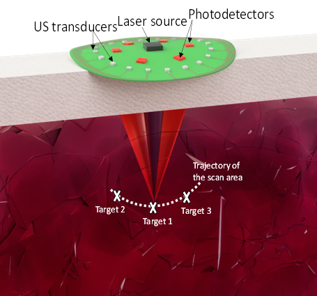
The researchers describe the method in a paper published in the journal Light: Science and Applications. Led by Maysam Chamanzar and Matteo Giuseppe Scopelliti, the team demonstrated using ultrasound to create a virtual "lens" within the body, rather than implanting a physical lens.
By using ultrasonic waves, they were able to effectively "focus" light within the tissue, which allows them to take images never before accessible non-invasively.
“Being able to relay images from organs, such as the brain, without the need to insert physical optical components will provide an important alternative to implanting invasive endoscopes in the body,” says Chamanzar. “We used ultrasound waves to sculpt a virtual optical relay lens within a given target medium, which for example, can be biological tissue. Therefore, the tissue is turned into a lens that helps us capture and relay the images of deeper structures.”
The researchers claim that In the future, the method can be implemented in the form of a handheld device or wearable surface patch, depending on the organ being imaged. Placing the device or patch on the skin, the clinician would be able to easily receive optical information from within the tissue to create images of what’s inside without endoscopy’s many discomforts and side effects.
The researchers think this new imaging technology could be applied in biomedical and clinical contexts within the next five years.
Source: Carnegie Mellon University press release


No comments:
Post a Comment